Description

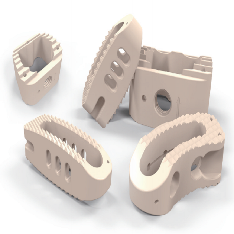
- Made of medical – grade PEEK material.
- Features a directional occlusal tooth design.
- Equipped with an extra – large bone graft window.
- Tantalum – metal marking rod for easy positioning.
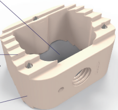
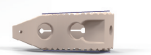
Cervical interbody fusion cage

Design of a large bone graft window:
It increases the integration rate and reduces the subsidence rate.
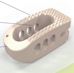
Design of directional occlusal teeth:
Effectively prevents the displacement of the intervertebral fusion cage between the vertebral bodies.
Its elastic modulus is similar to that of healthy bone tissue.
It is X-ray permeable.
It has better structural properties.

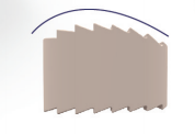
- It can effectively disperse the stress, maintain the balance between vertebral bodies, and restore the normal physiological lordosis of the cervical spine.
- When implanted, it can reduce the damage to the anterior edge of the vertebral body and effectively reduce the risk of prolapse.
Lumbar intervertebral fusion cage (implantation method of Posterior Lumbar Interbody Fusion, PLIF)

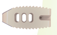
- Its elastic modulus is similar to that of healthy bone.
- It is X-ray permeable.
- It has better structural performance.
Three tantalum metal marking rods can determine the position of the intervertebral fusion cage under fluoroscopy.

It has a lordotic angle of 6°, which can restore the normal physiological lordotic curve.

The implantor can fully control and guide the implantation of the intervertebral fusion cage.
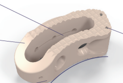
Schematic Diagram of the Steps of Using the Holding Device via the Transforaminal Lumbar Interbody Fusion (TLIF) Approach
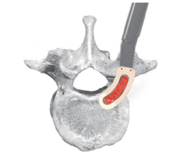
① After the endplate treatment is completed, firmly fix the intervertebral fusion cage to the head end of the holding device, and lock the locking knob at the tail end of the holding device. Then, implant it via the Transforaminal Lumbar Interbody Fusion (TLIF) approach.
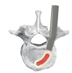
② When the tail end of the intervertebral fusion cage has completely entered the vertebral body, loosen the locking knob at the tail end of the holding device and continue the implantation.
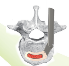
③ When the intervertebral fusion cage is close to the anterior edge of the vertebral body, it will automatically move forward in a horizontal position and finally be implanted in place at the horizontal position of the anterior edge of the vertebral body.
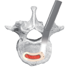
④ After confirming that the position of the fusion cage is correct, loosen the connecting part of the holding device, and finally remove the holding device to complete the implantation.